
 Spectral photon-counting X-ray · HR-GaAs:Cr
Science
For decades digital X-ray has relied on a scintillator and photocell pair. That method has reached the physical limits of spatial, contrast and time resolution. More than EUR 10 billion has been spent over 25 to 30 years looking for a way forward. Zeelta’s answer is a direct-conversion HR-GaAs:Cr sensor that counts photons and measures their energy.
01 · The limit
Why scintillator detectors cannot see more
In a scintillator, an absorbed X-ray quantum produces a flash of light that spreads in all directions. Only part of it reaches the photocells, and several cells are lit at once. Nearby events merge into one larger event. The detector cannot localise the event precisely and cannot measure the energy of the quantum. Its detective quantum efficiency (DQE) is about 35 to 40%.
Three ways out were studied. Dual-energy systems with two exposures did not add significant diagnostic information. Mathematical processing improved decision support but cannot go further without more data. The promising direction is a new detector base: monolithic direct-conversion semiconductor sensors such as a-Se, CdTe, CdZnTe and GaAs:Cr.
02 · Direct conversion
Photon counting with HR-GaAs:Cr
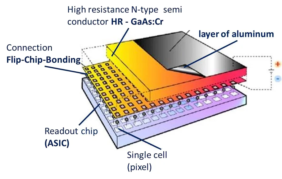
In a monolithic semiconductor sensor, an X-ray quantum creates an amount of charge proportional to its energy. Under the bias voltage the charges drift in opposite directions and are collected on a single pixel electrode. A readout ASIC amplifies the pulse, compares it with energy thresholds and counts it.
- The coordinate of each event is defined unambiguously; spatial resolution follows the pixel pitch, from 35 µm
- The charge gives the energy of the quantum: energy dispersion, the analogue of colour vision
- Two close simultaneous events are recorded separately
- DQE above 90%: quantum counting mode becomes possible
03 · Comparison
Scintillator + Si versus HR-GaAs:Cr
| Spatial resolution | Spectral (colour) | Density resolution | |
|---|---|---|---|
| Scintillator + Si | Pixel pitch 110 µm at best; actual resolution from 330 µm | Not available. Dual-energy methods need repeated exposures or two sources | 256 grey levels in routine radiography, 512 in expert mammography, 4,960 in 64-slice CT |
| HR-GaAs:Cr | Pixel pitch from 35 µm; actual resolution matches the sensor topology | Measures quantum energy accurately enough for qualitative and quantitative element analysis | About 4,000 grey levels, independently for each energy range |
| Can one replace the other? | Partially | No: spectral imaging is beyond scintillator systems | No: a separate grey scale per energy channel is beyond scintillator systems |
04 · Materials
The technological landscape
Monolithic semiconductor sensors suitable for channel-by-channel readout ASICs:
| Material | Strengths | Limitations |
|---|---|---|
| Silicon (Si) | Very cheap | Effective only at very low energies not used in medicine; fast radiation degradation |
| Amorphous selenium (a-Se) | First material to enable quantum counting | Needs thermal stabilisation and a horizontal panel; not used in real systems |
| CdTe, CdZnTe (CZT) | High efficiency at 10 to 120 keV, durable | About 293 USD per gram; panels up to 84.5 × 28.16 mm |
| HR-GaAs:Cr | DQE above 95% at 40 keV and about 75% at 120 keV; up to 70 °C; radiation resistance up to 10 GGy | About 7.2 USD per gram; flat panels larger than 430 × 430 mm |
05 · Results
What spectral X-ray shows
06 · Medicine
New methods of diagnostics
Spectral X-ray enables functional diagnosis, not only anatomical images. Research directions where we look for clinical partners:
- Vital dosimetry in real time (VDRT)
- A new cone beam CT system
- X-ray imaging and evaluation of the ocular vitreous body
- Pre-clinical testing and joint research with medical teams